
However, mild cases should still be monitored. If intermittent lameness becomes more frequent, the dog develops joint pain, or the kneecap slides out more often, the treatment plan may need to change.
Do Certain Dog Breeds Have Higher Risk for Patella Luxation Dog?

Yes. Small breed dogs and toy breeds are most often affected, especially with medial patellar luxation. Boston and Yorkshire terriers, Chihuahuas, Pomeranians, and other small breeds are commonly diagnosed.
Large breed dogs can also develop patellar luxation, especially lateral patellar luxation. Larger breeds such as Great Danes and Irish Wolfhounds are increasingly diagnosed with the condition.
When should I schedule a consultation at Bushnell Animal Clinic?
Schedule a consultation if your dog skips on a rear leg, limps, kicks the leg backward, yelps during movement, avoids stairs, refuses to jump, or shows a bow-legged or knock-kneed stance. You should also schedule a visit if symptoms appear only occasionally, because early patellar luxation may be intermittent.
A prompt exam can help determine whether the problem is mild and manageable or whether surgical treatment should be considered before further injury occurs.
What follow-up care is needed after treatment?
Follow-up care depends on whether your dog receives conservative management or surgery. Conservative care may include rechecks, weight monitoring, medication adjustments, joint supplements, and physical therapy exercises.
After patellar luxation surgery, follow-up care commonly includes restricted activity, incision checks, pain control, physiotherapy or exercise modification, and recheck visits with imaging when needed. Regular follow-up with the veterinary surgeon is essential to confirm that the knee is healing properly.
How do I know if my dog needs surgery versus conservative treatment?
The decision depends on the grade of patellar luxation, frequency of lameness, pain level, skeletal alignment, arthritis, age, activity level, and whether one or both knees are affected. Mild Grades 1 and 2 may be managed conservatively when symptoms are minimal.
Surgical correction is more often recommended for symptomatic Grade 2, Grade 3, and Grade 4 patellar luxation, especially when the dog has persistent lameness, significant clinical signs, or structural abnormalities that prevent the patella from staying in the normal groove. A veterinary exam and X-rays help determine the safest and most effective plan.
Introduction
Patellar luxation in dogs means the kneecap, or patella, slides out of its normal groove in the thigh bone and disrupts normal movement of the dog’s knee.

If your dog suddenly skips on a rear leg, limps for a few steps, kicks the affected leg backward, or avoids jumping, a luxating patella should be evaluated by a veterinarian.
This guide explains patella luxation dog symptoms, diagnosis, treatment options, surgery, recovery expectations, and cost considerations for pet parents in Bushnell, Sumter County, and surrounding Central Florida communities. It is written for dog owners who want to understand what is happening inside the knee joint, when conservative care may be enough, and when patellar luxation surgery may be the better long-term choice.
Patellar luxation occurs when the kneecap slides out of its normal position, causing intermittent lameness, joint pain, difficulty extending the knee, and sometimes progressive orthopedic changes. Because dogs luxating patella can have one or both knees involved, veterinary evaluation is important even when symptoms appear mild or come and go.
You will learn:
- How to recognize the classic “skipping” gait and other clinical signs
- What medial patellar luxation and lateral patellar luxation mean
- How veterinarians grade patellar luxation from Grade 1 through Grade 4
- When weight control, joint supplements, and physical therapy may help
- When surgical correction, including tibial tuberosity transposition, may be recommended
- What recovery, cost, and long-term prevention can look like in the Sumter County area
Patellar luxation in dogs means the kneecap, or patella, slides out of its normal groove in the thigh bone and disrupts normal movement of the dog’s knee. If your dog suddenly skips on a rear leg, limps for a few steps, kicks the affected leg backward, or avoids jumping, a luxating patella should be evaluated by a veterinarian.
This guide explains patella luxation dog symptoms, diagnosis, treatment options, surgery, recovery expectations, and cost considerations for pet parents in Bushnell, Sumter County, and surrounding Central Florida communities. It is written for dog owners who want to understand what is happening inside the knee joint, when conservative care may be enough, and when patellar luxation surgery may be the better long-term choice.
Patellar luxation occurs when the kneecap slides out of its normal position, causing intermittent lameness, joint pain, difficulty extending the knee, and sometimes progressive orthopedic changes. Because dogs luxating patella can have one or both knees involved, veterinary evaluation is important even when symptoms appear mild or come and go.
You will learn:
- How to recognize the classic “skipping” gait and other clinical signs
- What medial patellar luxation and lateral patellar luxation mean
- How veterinarians grade patellar luxation from Grade 1 through Grade 4
- When weight control, joint supplements, and physical therapy may help
- When surgical correction, including tibial tuberosity transposition, may be recommended
- What recovery, cost, and long-term prevention can look like in the Sumter County area
Understanding Patellar Luxation

Understanding how the patella, patellar tendon, patellar ligament, thigh bone, and shin bone work together helps explain why early diagnosis matters. In many affected dogs, patellar luxation results from developmental misalignment of the knee joint rather than a single injury, so signs may begin subtly and worsen as the dog grows or ages.
Be vigilant about the potential signs of patellar luxation in dogs, as they may indicate a need for veterinary evaluation.
What is Patellar Luxation
Patellar luxation is a kneecap dislocation in which the patella moves out of the trochlear groove, also called the femoral groove, at the front of the dog’s knee. The patella is a small bone that normally rides in this normal groove while the quadriceps muscle, patellar tendon, and patellar ligament help the knee extend properly.
In a healthy knee joint, the kneecap glides smoothly as the dog walks, runs, climbs stairs, or jumps. When the groove is too shallow, the long bones are misaligned, the soft tissues are unbalanced, or the pull of the quadriceps mechanism is abnormal, the patella slides out of place.
Patella luxation can cause discomfort and prevent the knee from extending properly. The affected limb may suddenly lock, skip, or lift off the ground until the knee cap returns to its normal position. In mild cases, the dog may seem normal between episodes; in severe patellar luxation, the affected knee may stay out of alignment much of the time.
Types of Patellar Luxation
There are two main directions a luxating patella can move. Medial patellar luxation, also called medial luxating patella, means the kneecap moves toward the inside of the leg. This is the most common form of luxating patella in dogs and is especially common in toy and small breed dogs.
Lateral patellar luxation means the kneecap moves toward the outside of the leg. This type is less common overall but is seen more often in large breed dogs and giant breeds. When the kneecap moves outward, the condition may be called lateral patellar luxation.
Patella luxation most frequently affects the hind legs of toy and small breed dogs, but can occur in larger breeds as well. Patellar luxation is primarily of genetic origin and is often a result of selective breeding for certain physical conformations, particularly in small and miniature dog breeds. Certain breeds, such as Boston and Yorkshire terriers, Chihuahuas, and Pomeranians, are more commonly affected by patellar luxation, while larger breeds like Great Danes and Irish Wolfhounds are also increasingly diagnosed with the condition.
The condition can be caused by a combination of genetic predisposition and developmental misalignment of the knee joint, leading to a shallow femoral groove that fails to properly accommodate the patella. Traumatic injuries can also lead to patellar luxation, but the majority of cases are non-traumatic and linked to congenital or developmental issues in the dog’s skeletal structure.
Grading System for Severity
Veterinarians categorize patella luxation into four distinct stages. There are four grades of patellar luxation, with higher grades indicating more severe conditions; approximately 50% of affected dogs may have both knees involved.
- Grade 1: The patella can be manually moved out of place during a physical exam, but it returns to the normal position on its own. Many dogs show few or no significant clinical signs.
- Grade 2: The patella slides out of place intermittently and may stay luxated until the dog extends the leg, kicks the back leg, or the veterinarian replaces it. This grade often causes intermittent lameness.
- Grade 3: The patella rides outside the normal groove most of the time but can be manually returned to position. The kneecap often slips back out again, and affected dogs may have more consistent lameness.
- Grade 4: The patella is permanently out of place and cannot be manually reduced. Severe patellar luxation may involve abnormally shaped femurs, tibial rotation, shallow trochlear groove anatomy, and obvious limb deformity.
When considering treatment options, it is crucial to discuss the specifics of patellar luxation in dogs with your vet.
Grading helps guide treatment decisions at Bushnell Animal Clinic, but grade is not the only factor. The dog’s condition, age, weight, pain level, arthritis, activity level, and whether one or both knees are affected all influence whether conservative care, knee surgery, or referral to a veterinary surgeon is most appropriate.
Recognizing and Diagnosing Patellar Luxation
Once you understand how the kneecap should move, the signs of patellar luxation become easier to recognize. Some dogs show only a brief skipping step, while others develop ongoing joint pain, reluctance to move, muscle loss in the affected leg, or abnormal posture.
Clinical Signs and Symptoms
Symptoms of patellar luxation can include a characteristic “skipping” lameness, where the dog may limp for a few steps and then return to a normal gait. Many pet parents describe seeing the dog pick up a rear leg, hop briefly, shake or kick the affected limb, and then walk normally after the kneecap pops back into place.
Signs of discomfort may include a dog yelping suddenly when the kneecap pops out, or showing reluctance to jump or run. Some dogs avoid stairs, hesitate before getting into a car, sit with one back leg angled out, or tire quickly during play.
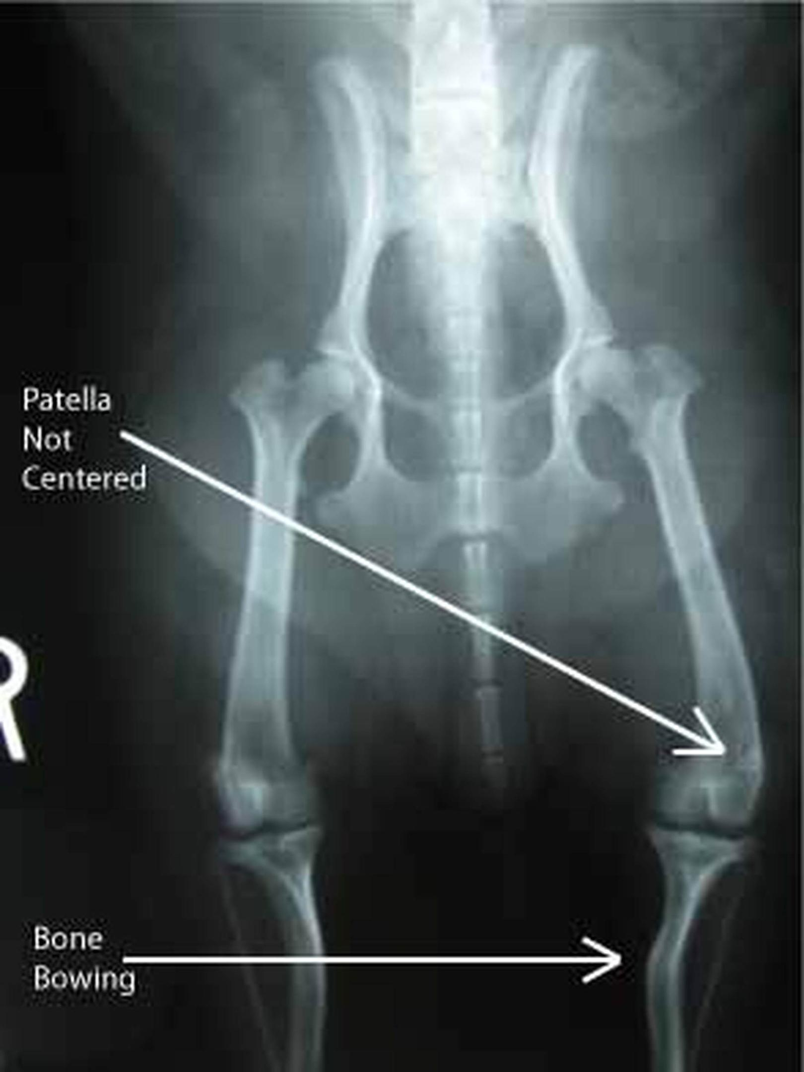
Mild luxating patella in dogs may cause occasional intermittent lameness with long normal periods between episodes. More severe cases can cause persistent limping, swelling around the affected knee, reduced ability to extend properly, and muscle loss in the hind limbs. In more severe cases of patellar luxation in dogs, dogs may develop abnormal posture such as bow-legged or knock-kneed stances.
A comprehensive understanding of patellar luxation in dogs can greatly assist pet owners in making informed decisions.
When to Seek Veterinary Care
Schedule an examination if your dog skips on a back leg, repeatedly shakes the rear leg, cries out when moving, avoids jumping, or develops any lameness that lasts more than a brief moment. Even if the dog run pattern returns to normal after a few steps, the kneecap may still be unstable.
Without treatment or monitoring, symptoms can progress over time. Repeated kneecap dislocation may irritate the joint capsule, stretch soft tissues, worsen cartilage wear, and contribute to arthritis. Dogs with Grade 2 patellar luxation may appear mildly affected at first but later develop more frequent lameness.
Early intervention gives your veterinarian more options. Mild cases may respond to weight control, joint supplements, exercise modification, and physical therapy, while more advanced cases may need surgical repair before further injury or major degenerative change occurs.
Diagnostic Procedures
Patellar luxation diagnosed through a physical examination involves the veterinarian palpating the knee to detect abnormal movement of the kneecap, and X-rays may be used to assess the severity and any associated damage. During the exam, Dr. Roger Hart at Bushnell Animal Clinic may evaluate gait, flex and extend the dog’s knee, check whether the patella moves inward or outward, and assess both knees because bilateral involvement is common.
A routine physical examination can often identify patellar luxation in dogs and whether the problem is called medial patellar luxation or lateral patellar luxation. The physical exam also helps distinguish patella luxation from other orthopedic conditions, such as cranial cruciate ligament injury, hip dysplasia, fractures, or soft tissue strains.
X-ray imaging helps evaluate the trochlear groove, thigh bone, shin bone, limb alignment, arthritis, and whether surgical procedures may be needed. In complex cases, large dogs, severe patellar luxation, or dogs with suspected angular limb deformities may need advanced imaging or referral for surgical planning.
Treatment Options and Recovery
Treatment for luxating patella in dogs should be individualized. At Bushnell Animal Clinic, the goal is to match the treatment plan to the grade of luxation, the dog’s pain level, the knees involved, the owner’s ability to manage recovery, and the long-term quality of life expected for the patient, drawing on a full range of veterinary services for dogs in Bushnell.
Conservative Management
Mild cases of patella luxation, especially Grades 1 and 2, can often be managed with weight control, joint supplements, or physical therapy. Conservative care does not reshape the femoral groove or surgically correct bone alignment, but it can reduce stress on the affected knee and help many dogs stay comfortable.
Weight management is one of the most important steps. Keeping dogs at a healthy weight reduces load on the knee joint, limits joint pain, and may slow the progression of arthritis. This is especially important in Central Florida, where year-round warm weather can encourage activity but also makes overheating a concern for overweight dogs.
Exercise modification may include avoiding repeated jumping, limiting stairs, using ramps, choosing leash walks over rough play, and encouraging low-impact strengthening. Physical therapy may include controlled range-of-motion exercises, muscle-building work for the quadriceps muscle, balance exercises, and in some cases hydrotherapy or underwater treadmill work.
Pain management may involve veterinarian-prescribed anti-inflammatory medication, adjunctive pain control, and joint supplements. Conservative management is most appropriate when clinical signs are mild, the patella returns to normal position easily, and the dog is not developing worsening lameness or structural change.
Surgical Treatment Procedures
Understanding the risks associated with patellar luxation in dogs can help in early prevention and treatment strategies.
Surgery becomes more likely when a dog has repeated lameness, joint pain, Grade 2 disease that is worsening, or Grade 3 to Grade 4 patellar luxation. Surgical treatment for patellar luxation is often recommended for grades II through IV, and the costs can vary significantly based on the complexity of the procedure and the dog’s condition.
Surgical treatment for patellar luxation often involves procedures such as tibial tuberosity transposition, trochlear wedge resection, and lateral capsular imbrication to restore normal knee joint anatomy, with the kneecap and limb alignment surgically corrected through these techniques. Tibial tuberosity transposition moves the attachment point on the shin bone so the patellar tendon pulls the kneecap in better alignment. Trochlear groove deepening techniques, including trochlear wedge resection or recession sulcoplasty, create a deeper track for the patella.
Soft tissue reconstruction may tighten loose tissues, release tight tissues, or reinforce the joint capsule so the patella stays centered. Surgical options for patellar luxation may include femoral varus osteotomy, recession sulcoplasty, and soft tissue reconstruction, depending on the severity and specific characteristics of the condition. In dogs with more severe bone deformity, femoral or tibial osteotomy may be needed to correct abnormal alignment of the long bones.
After surgery for patellar luxation, dogs typically require a recovery period of at least eight weeks, during which regular follow-up visits with the veterinary surgeon are essential to monitor progress. Recovery from patellar luxation surgery typically requires a period of exercise restriction lasting at least six weeks, followed by a gradual return to normal activity as advised by the veterinarian.
Postoperative care for dogs recovering from patellar luxation surgery often includes limiting physical activity and may involve a structured rehabilitation plan that includes physiotherapy and exercise modification. Most dogs need leash walks only, no running or jumping, pain medication as prescribed, incision monitoring, and follow-up imaging when recommended to confirm bones heal appropriately.
The most common postoperative complication in patellar luxation surgery is the recurrence of the luxating patella, along with risks of implant failure, under- or overcorrection, and infections. Careful surgical planning, strict activity restriction, weight control, and follow-up visits help reduce these risks and support a smoother dog recover process.
Treatment Comparison
Criterion | Conservative Management | Surgical Treatment |
|---|---|---|
Grade of luxation | Often appropriate for Grade 1 and some mild Grade 2 cases | Often recommended for symptomatic Grade 2, Grade 3, and Grade 4 cases |
Expected outcomes | May reduce pain and improve function but does not correct abnormal anatomy | Aims to restore alignment and keep the patella in the normal groove |
Recovery time | Ongoing management with gradual strengthening and monitoring | At least six weeks of exercise restriction; often at least eight weeks of structured recovery and follow-up |
Cost considerations | Lower upfront cost; may include exams, X-rays, medication, supplements, and rehab | Higher upfront cost; varies by dog size, severity, implants, and whether one or both knees are treated |
Long-term prognosis | Good for many mild cases if weight and activity are managed | Generally favorable when surgical correction is appropriate and recovery instructions are followed |
The right choice depends on the dog’s condition, not just the grade. A small dog with mild intermittent lameness may do well with conservative management, while a young active dog with frequent patella slides may benefit from corrective surgery before arthritis progresses. Most dogs with severe patellar luxation need surgical evaluation because conservative care cannot correct major structural abnormalities. |
Patellar luxation in dogs is common, especially in toy and small breed dogs, but it can affect large breed dogs as well. The key is early recognition: a skipping gait, intermittent lameness, yelping when the kneecap pops, reluctance to jump, or abnormal rear-leg posture should not be ignored in cases of patellar luxation in dogs.
Pet parents often worry about pain, cost, recurrence, and how to keep an active dog quiet long enough to heal. These concerns are valid, especially when the affected dog is young, energetic, or has both knees involved.
Managing Recovery Complications
Post-surgical swelling, bruising, mild discomfort, and temporary stiffness can occur after patellar luxation surgery. Follow your veterinarian’s instructions for medication, incision care, bandage care if used, and scheduled rechecks.
The biggest home-care challenge is activity restriction. Use a crate, small room, baby gates, leash walks, non-slip flooring, and ramps to prevent jumping. If the dog is too active, ask your veterinarian whether calming strategies or medication support are appropriate during the early healing period.
Watch for warning signs such as worsening swelling, discharge, sudden non-weight-bearing lameness, fever, chewing at the incision, or pain that does not improve. These signs need prompt veterinary attention because infection, implant irritation, delayed healing, or recurrence can affect the outcome.
Preventing Re-injury
Long-term success depends on protecting the knee joint after recovery. Dogs should return to activity gradually, with controlled leash walks first and higher-impact activity only when cleared by the veterinarian.
Maintain muscle strength with regular low-impact exercise. Avoid repetitive jumping from couches, trucks, beds, or porches, especially for small breed dogs with a history of medial patellar luxation. For large breed dogs or large dogs with lateral patellar luxation, controlled conditioning is just as important because added force across the knee can stress healing tissues.
Weight management is crucial for dogs recovering from patellar luxation surgery, as maintaining a healthy weight can help reduce stress on the knee joint and slow the progression of arthritis. For Sumter County pet owners, this may include measured meals, regular weigh-ins, heat-safe walking schedules, and joint-friendly exercise during cooler parts of the day.
Cost and Financial Planning
The cost of luxating patella surgery in dogs can range from $1,000 to $5,000 per leg, depending on factors such as the dog’s size, the severity of the luxation, and whether one or both knees are affected. In Central Florida, pricing may also vary based on X-rays, bloodwork, anesthesia, implants, referral to a veterinary surgeon, overnight care, physical therapy, and whether advanced surgical procedures are required.
At Bushnell Animal Clinic, cost transparency begins with diagnosis and a written treatment plan whenever possible. A Grade 1 or mild Grade 2 case may involve exam fees, imaging, medication, joint supplements, and rehab planning, while severe patellar luxation may require surgical repair, implants, follow-up X-rays, and a longer recovery plan; similar planning applies to other conditions such as canine cryptorchidism and related surgical care.
Pet insurance can help cover the costs associated with luxating patella treatment, including surgery, with some policies offering reimbursement options up to 90%. However, many policies exclude pre-existing conditions or have orthopedic waiting periods, so pet parents should review coverage before symptoms begin when possible.
Conclusion and Next Steps
If your dog is diagnosed with patellar luxation in dogs, it’s essential to understand the condition and the potential treatments available.Patellar luxation in dogs is common, especially in toy and small breed dogs, but it can affect large breed dogs as well. The key is early recognition: a skipping gait, intermittent lameness, yelping when the kneecap pops, reluctance to jump, or abnormal rear-leg posture should not be ignored.
Being proactive about patellar luxation in dogs can help mitigate its effects on your pet’s quality of life.
If you suspect a luxating patella, take these next steps, starting with reaching out through the clinic’s contact information for Bushnell Animal Clinic:
- Schedule a veterinary examination to have the affected knee and the opposite knee evaluated.
- Write down symptoms such as skipping, limping, kicking the leg, or reluctance to run or jump.
- Bring medical history including prior injuries, medications, supplements, and changes in activity.
- Ask about grading and imaging so you understand whether the patella luxation is mild, moderate, or severe.
- Discuss conservative care versus surgery based on your dog’s grade, pain, age, weight, and long-term needs.
Related orthopedic concerns, including hip dysplasia, cruciate ligament injury, arthritis, and other causes of hind-limb lameness, may require similar diagnostic attention. Bushnell Animal Clinic is committed to helping Sumter County families understand their options and choose comprehensive, affordable care for each dog’s condition.
Frequently Asked Questions
Can patellar luxation be prevented in puppies?
Patellar luxation in dogs cannot always be prevented because many cases are genetic or developmental. Patellar luxation in dogs is primarily of genetic origin and is often linked to selective breeding for certain physical conformations, particularly in small and miniature dog breeds.
Risk can be reduced by responsible breeding, screening high-risk breeds, keeping puppies at a healthy weight, avoiding excessive strain during growth, and scheduling early veterinary exams if a puppy skips, limps, or has abnormal hind-limb posture related to patellar luxation in dogs.
How much does patellar luxation surgery cost in Central Florida?
Luxating patella surgery can range from $1,000 to $5,000 per leg, depending on the dog’s size, severity of the luxation, complexity of the procedure, and whether one or both knees are affected. Costs in the Sumter County and Central Florida area may vary depending on diagnostics, implants, surgical time, follow-up imaging, and referral needs.
Being educated about patellar luxation in dogs can empower pet owners to take proactive steps for their pets’ health.
A personalized estimate is the best way to understand cost for your dog because a mild medial patellar luxation in a small dog is very different from severe patellar luxation in a large dog with bone deformity.
What is the recovery time for patellar luxation surgery?
After surgery for patellar luxation, dogs typically require at least eight weeks of recovery, with regular follow-up visits to monitor healing. Exercise restriction usually lasts at least six weeks, followed by a gradual return to normal activity as advised by the veterinarian.
Understanding treatment options for patellar luxation in dogs is essential for responsible pet ownership.
Regular check-ups can help in the early diagnosis of patellar luxation in dogs, allowing for better management.
Recognizing the signs of patellar luxation in dogs allows for timely veterinary intervention.
The importance of regular veterinary checks cannot be stressed enough for patellar luxation in dogs.
Some dogs regain comfortable function within a few months, while others need longer physical therapy, especially if arthritis, severe deformity, or both knees are involved.
Can dogs live normal lives with mild patellar luxation?
Yes. Many dogs with Grade 1 or mild Grade 2 patellar luxation live comfortable, active lives with weight control, exercise modification, joint supplements, and physical therapy.
However, mild cases should still be monitored. If intermittent lameness becomes more frequent, the dog develops joint pain, or the kneecap slides out more often, the treatment plan may need to change.
Pet owners should familiarize themselves with patellar luxation in dogs to ensure their pets receive appropriate care.
Understanding the signs of patellar luxation in dogs can help pet owners take timely action.
Do certain dog breeds have higher risk for patellar luxation?
Yes. Small breed dogs and toy breeds are most often affected, especially with medial patellar luxation. Boston and Yorkshire terriers, Chihuahuas, Pomeranians, and other small breeds are commonly diagnosed.
Large breed dogs can also develop patellar luxation, especially lateral patellar luxation. Larger breeds such as Great Danes and Irish Wolfhounds are increasingly diagnosed with the condition.
When should I schedule a consultation at Bushnell Animal Clinic?
Schedule a consultation if your dog skips on a rear leg, limps, kicks the leg backward, yelps during movement, avoids stairs, refuses to jump, or shows a bow-legged or knock-kneed stance. You should also schedule a visit if symptoms appear only occasionally, because early patellar luxation may be intermittent. Timely intervention is key when dealing with patellar luxation in dogs, ensuring the best possible outcome for your furry friend.
A prompt exam can help determine whether the problem is mild and manageable or whether surgical treatment should be considered before further injury occurs.
What follow-up care is needed after treatment?
Follow-up care depends on whether your dog receives conservative management or surgery. Conservative care may include rechecks, weight monitoring, medication adjustments, joint supplements, and physical therapy exercises.
After patellar luxation surgery, follow-up care commonly includes restricted activity, incision checks, pain control, physiotherapy or exercise modification, and recheck visits with imaging when needed. Regular follow-up with the veterinary surgeon is essential to confirm that the knee is healing properly.
How do I know if my dog needs surgery versus conservative treatment?
Moreover, early detection of patellar luxation in dogs can lead to better management and outcomes. The decision depends on the grade of patellar luxation, frequency of lameness, pain level, skeletal alignment, arthritis, age, activity level, and whether one or both knees are affected. Mild Grades 1 and 2 may be managed conservatively when symptoms are minimal.
Surgical correction is more often recommended for symptomatic Grade 2, Grade 3, and Grade 4 patellar luxation, especially when the dog has persistent lameness, significant clinical signs, or structural abnormalities that prevent the patella from staying in the normal groove. A veterinary exam and X-rays help determine the safest and most effective plan.
Leave a Reply